
What digital scanning is
In dentistry, digital scanning usually means an intraoral scan. A small handheld scanner is moved around your mouth, and it records your teeth and gums as a three-dimensional (3D) model on a computer. It is the digital alternative to a conventional impression, in which a tray of soft material is pressed over the teeth and left to set.
The scanner projects light onto the teeth and gums1, and software joins the images into a 3D surface model. It records surfaces only and does not use X-rays. A "3D scan" of the jaw bone (CBCT) is a different test: it is an X-ray.
On this page, "we" and "our clinic" mean our clinic in Antalya.
- An intraoral scan records the surfaces of your teeth and gums with light, as a 3D model on a computer. It is not an X-ray.
- In clinical studies, scanning was generally quicker than a conventional impression, and patients generally preferred it, though not in every study.
- Scans are not more accurate in every case. Edges under the gum and bleeding are hard to capture. For full-arch bridges on more than four implants, a 2025 review found conventional impressions slightly more accurate than digital methods taken together.
- A 2025 review advised against choosing the shade of a crown or veneer with a scanner, although its evidence was of very low certainty.
- Ask for copies of your scan files, in an open format and in the original one, and ask how long they are kept.
Common uses in dentistry
Intraoral scanners are used for diagnosis and for making restorations or custom devices1 in restorative dentistry, implant work and orthodontics. A 2017 review of 132 studies describes these uses, among others:
- Crowns, bridges, inlays and onlays. The scan records the prepared tooth, its neighbours and the opposing teeth. Our dental crowns page explains what a crown involves.
- Crowns and bridges on implants. A small cylinder called a scan body is screwed onto each implant, so that the scan can record the 3D position of the implant1. See our dental implant treatment page.
- Planning implant surgery. A scan of the teeth and gums can be combined with a CBCT scan1 of the bone. Together they are used to plan where implants go and to design a surgical guide.
- Orthodontics. Scans are used for diagnosis and planning, and as the starting point for aligners and other custom-made appliances1.
- Records and explanation. A 3D model can be kept as a record of your mouth, and it can help the dentist show you what they see.
- Smile design, posts and cores (built into a root-filled tooth to hold a crown) and partial dentures, according to the same review. It found only a few studies on scans for partial or complete dentures1.
For complete dentures, scanning a mouth with no teeth is harder; see "When conventional impressions may be used" below.
How an intraoral scan is captured
Scanners and techniques differ, so the details vary. The outline below is typical for a crown or bridge, and the model builds up on a screen as the scan goes on.
Older scanners needed a thin powder on the teeth. More recent ones can scan without powder1. You keep your mouth open while the tip of the scanner is moved over your teeth, including the back teeth. The size of the tip varies between scanners, and a smaller tip is preferable for comfort1. If part of the scan is unclear, that part can be deleted and captured again without repeating the entire procedure1.
Preparing the area
The teeth to be scanned are cleaned, and any bleeding is controlled. For a tooth prepared for a crown, the dentist may first place a thin cord in the narrow gap between the tooth and the gum. This helps to show the edge of the preparation.
Scanning each jaw
The scanner tip is moved slowly over the teeth and gums. The software joins the images into a 3D model of the jaw. Both jaws are usually scanned.
Recording the bite
The teeth are usually scanned while you bite together, so that the model shows how the upper and lower teeth meet.
For implants: scan bodies
For a restoration on implants, a scan body is screwed onto each implant it will sit on, and scanned. This records the position of those implants.
Checking the model
The dentist checks the model on the screen and rescans any missing or unclear area before the file is used.
Scan review and data quality
A scan can only record what the scanner can see. The dentist checks the model before it is used. A 2017 review notes that the dentist and the dental technician can assess the quality of the scan in real time1. If the file reaches the laboratory while you are still there, the technician can ask straight away for another scan1. A problem found later may mean another visit.
What affects the quality of a scan
- Edges under the gum. Where the edge of a prepared tooth lies under the gum, it can be hard to capture. Unlike impression material, light cannot physically push the gum aside1. A thin cord placed between the tooth and the gum helps to show the edge.
- Bleeding. Blood may hide the edges1 of the preparation.
- Gum health. As with conventional impressions, healthy soft tissues are essential for a good scan1.
- Experience and technique. There is a learning curve1 for the person scanning. It is also still unclear whether one scanning strategy is better than another1.
- The scanner. Studies, almost all in the laboratory, found that different scanners have different accuracy1.
Colour and shade
Some scanners record colour. The 2017 review sees colour information as useful mainly for communication with the patient1, and of less clinical importance. For the shade of a crown or veneer, a 2025 meta-analysis compared scanners with a measuring instrument (a spectrophotometer). A shade tab is one sample tooth in a shade guide. The scanners picked the same tab as the instrument in about 38 in 100 readings with one guide, and about 28 in 100 with another2. Its authors do not recommend choosing shade with an intraoral scanner, although the certainty of this evidence was very low.
The shade is usually chosen with a shade guide, and the light in the room matters. In one laboratory study, dentists with normal colour vision matched shades better under a standard daylight lamp than under a tungsten bulb3. In another, handheld shade-matching lights and training both improved dental students' scores4. Our dental laboratory page explains how the shade reaches the laboratory.
Connection to planning and laboratory workflows
A scan becomes a digital file. With a conventional impression, a plaster model is usually poured before anything is made. With a scan, no plaster model is needed1, and the 3D model can be sent to the dental laboratory electronically1.
Working from the scan, the restoration is usually designed on a computer (CAD) and then made by machine, for example milled from a block (CAM). Some clinics design and mill restorations in the clinic itself1 from the scan; others send the file to a laboratory. Your dentist will tell you which applies to your treatment, and you can ask for it in writing. Our CAD/CAM page covers design, manufacture and what the evidence says about restorations made this way.
For implants, the scan of the teeth and gums can be superimposed on a CBCT scan1 of the bone. Together they show the teeth, the gum surface and the bone. They are used to plan the position of each implant and to design a surgical guide. Our digital treatment planning page covers planning software, guides and their limits.
The scan is a record, not a plan. A dentist still decides what the restoration or treatment should be, and the laboratory and the planning software work from that decision.
Benefits and patient experience
The findings below come from published studies, not from our own records.
- No impression tray. A scan avoids the tray of setting material. Some people, for example those with a strong gag reflex1, find a tray hard to tolerate.
- Usually quicker. A 2021 systematic review of 17 clinical studies in 437 patients found scanning overall faster than conventional impressions5 for crowns, bridges and implant work. The results were described study by study and not pooled. In one trial, a conventional technique was faster. Scans also needed more retakes, though the average retake time was lower5.
- Generally preferred by patients. In the same review, scanning was generally preferred5. The one trial in which the conventional technique was faster found no difference in comfort.
- Similar results for the restorations. The studies in that review that followed the restorations reported similar results with both methods5.
- Time saved after the scan. A 2017 review notes that most of the time saved comes afterwards, during the steps that follow1, not from the scan itself.
- Seeing your own mouth. With a model on the screen, patients can feel more involved in their treatment1, and the dentist can show you what they see.
What it is like
You need to keep your mouth open while the tip is moved around, and reaching the back teeth can be awkward. Tell the dentist if you need a break.
Accuracy limitations
Accuracy here means how closely the scan matches your mouth. It is measured mostly in laboratory studies, and it is not the same as how long a restoration lasts. The studies below compare methods in general. They do not show how accurate any particular scanner, or any particular clinic's scans, are.
- Single crowns and short bridges. The 2017 review judged scan accuracy clinically satisfactory and similar to that of conventional impressions1 for single teeth. It reached the same view for bridges of up to four or five units (each unit is one tooth of the bridge).
- Long bridges and full arches. The same review found that scans did not appear to have the same accuracy1 for long bridges and full-arch work, on teeth or implants. It also warned that newer scanners improve faster than studies can be published.
- Full-arch bridges on implants. A 2025 meta-analysis covered 37 studies, 30 of them laboratory studies. It found no significant overall difference in accuracy6 between digital and conventional impressions. Intraoral scanners on their own did not differ from conventional impressions6. With more than four implants, conventional impressions were slightly more accurate6 than digital methods taken together (intraoral scans and photogrammetry, a camera-based method). The authors suggest that accuracy may decline when scanning arches with more than four implants6. Finding no significant difference does not prove that the two methods are equal.
- Implants where some natural teeth remain. A 2025 review of seven clinical studies in 151 patients found digital impressions a clinically acceptable alternative for short-span implant restorations7. Digital impressions differed less from the reference implant positions on one measure, with no difference in angle, but the studies varied widely. The authors advise caution with long spans or angled implants7.
- Edges under the gum and bleeding. Both can stop the scanner recording the edge of a prepared tooth (see "Scan review and data quality" above).
- Shade. A 2025 review advised against choosing the shade with a scanner (see above).
| Intraoral scan | Conventional impression | |
|---|---|---|
| How the mouth is recorded | Light, as a 3D model on a computer | Soft material in a tray, then usually a plaster model |
| Tray of material in the mouth | No | Yes |
| Single crowns and short bridges | Accuracy similar in a 2017 review | Accuracy similar in a 2017 review |
| Full-arch bridge on more than four implants | Accuracy may decline, according to a 2025 review | Slightly more accurate than digital methods taken together, in the same review |
| Edges deep under the gum | Light cannot push the gum aside | The material can push the gum aside, but bleeding still has to be controlled |
| Getting it to the laboratory | Sent as a file | The impression or model is sent |
| If part of it is unclear | That part can be rescanned | Usually the impression is taken again |
When conventional impressions may be used
A conventional impression is still a normal choice, and neither method suits every case. Reasons it may be used include:
- Long bridges and full-arch work, especially on many implants (see "Accuracy limitations").
- Edges deep under the gum. Light cannot push the gum aside, and some authors suggest combining the two methods1, partly using impression material. With either method, healthy gums are essential1 and bleeding has to be controlled.
- Complete dentures. For a mouth with no teeth, scanning still presents some issues1. There are few fixed reference points, and the scan cannot record how the soft tissues move. See our dentures page.
- Angled implants or long spans in people who still have some teeth (see "Accuracy limitations").
- The equipment and the laboratory. Scanners differ in accuracy, and not every clinic or laboratory works with scans.
Your dentist will tell you which method is planned for you and why, and which scanner is used if your teeth are scanned. You can ask for this in writing before you decide.
Data handling and repeat scans
Your scan is a file
A scanner saves the model in its maker's own (proprietary) format, in an open format such as STL, or in both. The 2017 review stated that open-format files can be opened by all dental design (CAD) systems1. Files from some closed systems can be opened only with software from the same maker1, and converting files may lose quality and information1. If another dentist may need your files later, check with them which format they can open. Ask for a copy in an open format and in the original format too.
How scans are stored and shared
A scan is a record of your mouth. Ask before treatment:
- whether your scan will be sent to a laboratory or anyone else, and where that laboratory is;
- how the files are sent and stored, and who can see them;
- how long they are kept, and how you can get a copy later.
Private dental clinics in Turkey record the diagnosis, the treatment and any X-rays in detail, with tooth numbers8. You can examine your file and records and get a copy9. Ask whether your scan files are included, and ask for the copies in writing.
Repeat scans
A scan records your mouth on the day it is taken. If a tooth is prepared or treated, teeth move or the gum changes, an older scan may no longer match. A new one is then taken. Because the scanner uses light, repeating a scan adds no X-ray dose.
Questions and consent before treatment
Before treatment, you can ask:
- whether a scan or a conventional impression will be used, and why;
- which scanner is used, and whether the files can be exported in an open format such as STL;
- who takes and checks the scan, and who designs and makes the restoration, and where;
- what happens if a restoration does not fit, and who pays for a remake.
You have the right to be told, on request, who is treating you, and their role and title9. Ask for the answers in writing. Ask also for a written record of each restoration: the material, the product and the laboratory that made it.
Consent. Turkey's patient rights regulation says what you are told before treatment. It includes the other options, with their benefits and risks, and the possible consequences of refusing9. In private dental clinics, you are asked to sign a consent form for every intervention8. Two copies of the form are signed, and one is given to you9. Ask for your copy. You can say no, or ask to stop, at any stage. The scan itself removes nothing from your teeth, but once a tooth has been prepared (shaped) for a restoration, that step cannot be undone.
Frequently Asked Questions
Is an intraoral scan an X-ray?
No. An intraoral scanner records the surfaces of your teeth and gums with light. It does not show the bone or the roots. A CBCT "3D scan" of the jaw is a different test, which uses X-rays.
Is a digital scan more accurate than a mould?
Not in every case. For single crowns and short bridges, a review found scan accuracy similar to that of conventional impressions. For long bridges and full arches on many implants, a conventional impression may still be the better choice.
Is scanning more comfortable than an impression?
There is no tray of setting material, which helps people with a strong gag reflex. You keep your mouth open while the scanner tip is moved around. In studies, patients generally preferred scanning, though not in every study.
Can the scanner choose the shade of my crown?
A 2025 review advised against it, although the evidence was weak. The shade is usually chosen with a shade guide in good light, sometimes with photographs or a measuring instrument as well.
Can I get a copy of my scan?
Ask before treatment. You can examine your file and records and get a copy. Ask for the scan files in an open format, such as STL, and in the original format too. Ask how long the clinic keeps them, and who else receives them.
Why might I need to be scanned again?
An unclear part of a scan can be rescanned on its own. A new scan may also be needed when your mouth has changed, for example after a tooth is prepared or treated. The scanner uses light, so a repeat adds no X-ray dose.
Can a scan be used for dentures?
A 2017 review lists partial dentures among the uses of scanning, but found only a few studies on it. For complete dentures, scanning a mouth with no teeth is harder, so a conventional impression may be preferred.
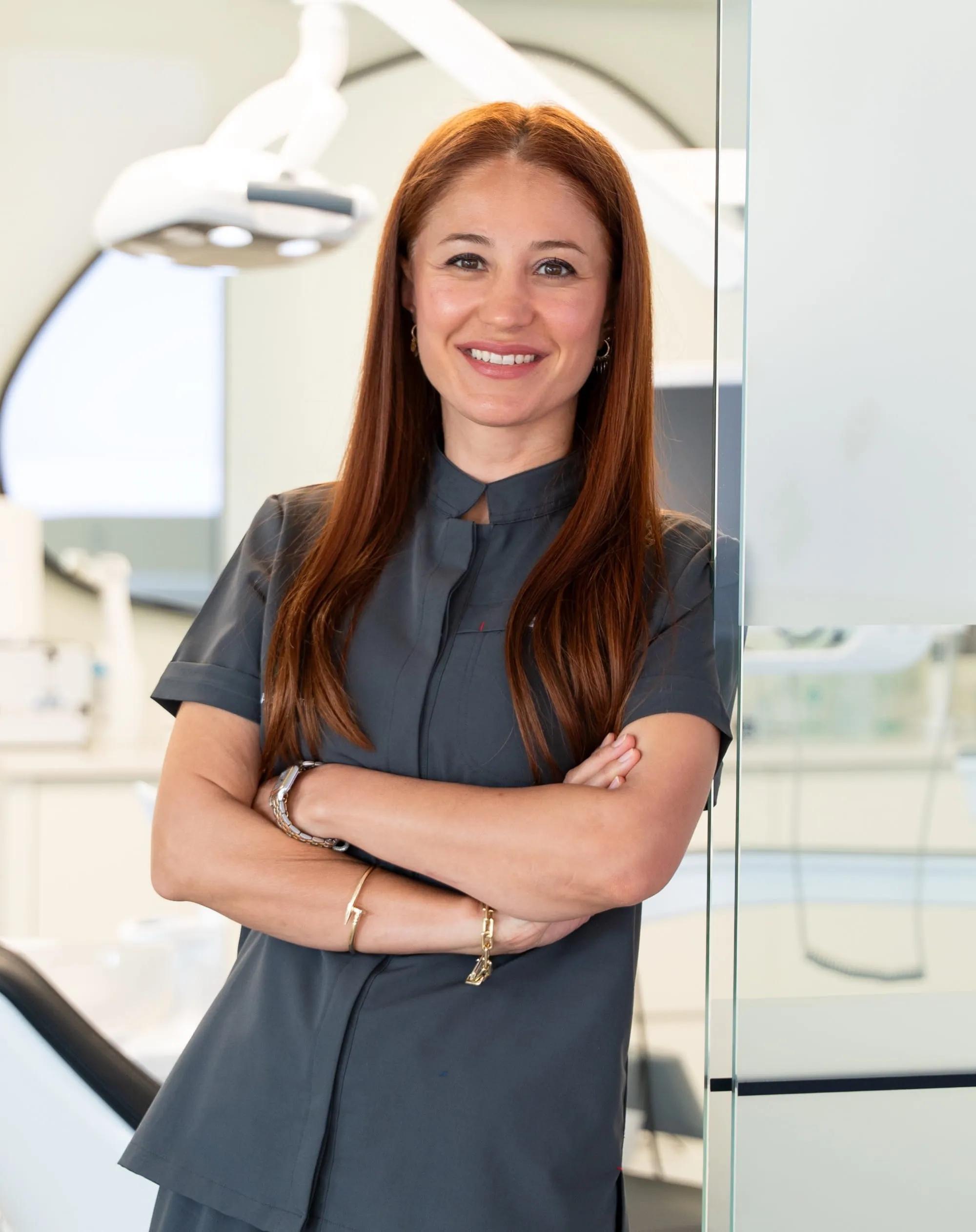
Dt. Dilek AKSU GÜLER
Dentist · Founder
She has completed advanced training in implantology and works in aesthetic restorations and smile design.
Sources
- Intraoral scanners in dentistry: a review of the current literature. BMC Oral Health 2017;17(1):149 (Mangano F, Gandolfi A, Luongo G, Logozzo S). 2017.↩doi.org
- Color comparison between intraoral scanner and spectrophotometer shade matching: a systematic review and meta-analysis. J Esthet Restor Dent 2025;37(2):361-377. 2024.↩doi.org
- Shade matching performance of normal and color vision-deficient dental professionals with standard daylight and tungsten illuminants. J Prosthet Dent 2010;103(3):139-47. 2010.↩doi.org
- Influence of light source, polarization, education, and training on shade matching quality. J Prosthet Dent 2016;116(1):91-7. 2016.↩doi.org
- Intraoral scanning reduces procedure time and improves patient comfort in fixed prosthodontics and implant dentistry: a systematic review. Clin Oral Investig 2021;25(12):6517-6531 (Siqueira R et al.). 2021.↩doi.org
- Comparative analyses of accuracy between digital and conventional impressions for complete-arch implant-supported fixed dental prostheses: a systematic review and meta-analysis. J Prosthodont 2026;35(3):252-274 (Alfaraj A et al.). 2025.↩doi.org
- Accuracy of digital versus conventional implant impressions in partially dentate patients: a systematic review and meta-analysis. Journal of Dentistry 2025;160:105918 (Park JS, Alshehri YFA, Kruger E, Villata L). 2025.↩doi.org
- Ağız ve Diş Sağlığı Hizmeti Sunulan Özel Sağlık Kuruluşları Hakkında Yönetmelik (consolidated text). T.C. Sağlık Bakanlığı; Resmî Gazete 6/10/2022 No 31975, amended RG 5/3/2024-32480 and 15/12/2024-32753. 2022.↩mevzuat.gov.tr
- Hasta Hakları Yönetmeliği (consolidated text). T.C. Sağlık Bakanlığı; Resmî Gazete 1/8/1998 No 23420, amended RG 8/5/2014-28994, 23/12/2016-29927, 16/1/2019-30657. 1998.↩mevzuat.gov.tr